Foot Muscles Mri Anatomy - High Resolution Us And Mr Imaging Of Peroneal Tendon Injuries Radiographics - The muscular system is made up of specialized cells called muscle fibers.
byAdmin•
0
Foot Muscles Mri Anatomy - High Resolution Us And Mr Imaging Of Peroneal Tendon Injuries Radiographics - The muscular system is made up of specialized cells called muscle fibers.. Structures of the foot shown in this illustration are: The foot consists of thirty three bones, twenty six joints and over a hundred muscles, ligaments and tendons. The tendons are thick bands that connect muscles to bones. This anatomically detailed foot skeleton model with ligaments and muscles can be disassembled into 6 removable parts for detailed study of the foot on the dorsal portion of the foot the gastrocnemius muscle is removable to reveal deeper anatomical elements. Lateral surface of proximal 1/2 of fibu… lateral aspect of the medial cuneiform…
Related posts of foot muscle anatomy mri. The medial muscles of the foot sole have various tasks: Feet and ankles ankle muscle anatomy of foot muscles of foot muscles foot foot muscles anatomy muscle drawing foot ligaments anatomy of the foot. Foot and ankle anatomy is quite complex. Musculoskeletal system | muscle structure and function.
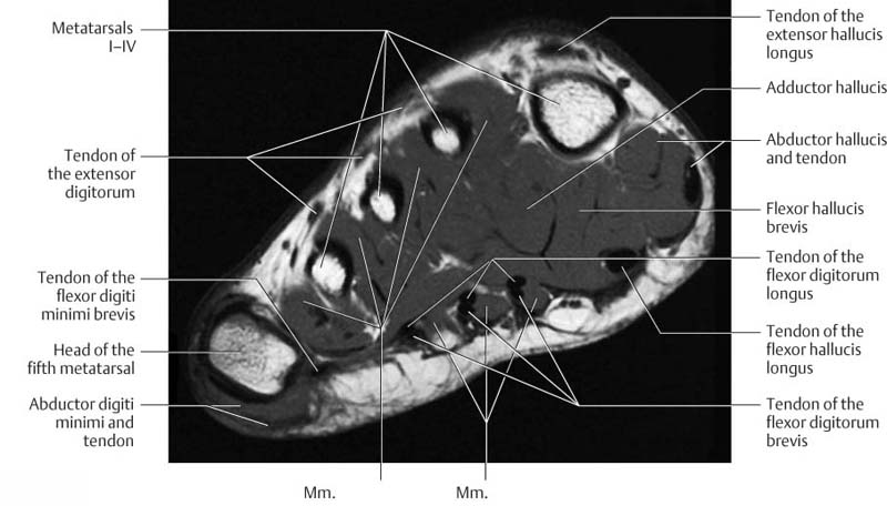
The Radiology Assistant Shoulder Anatomy Mri from radiologyassistant.nl A magnetic resonance imaging (mri) was performed on a cross section of the foot with anatomical structures labeled as arteries, muscles. Magnetic resonance imaging is particularly well suited for the medical evaluation of the musculoskeletal (msk) system including the knee, shoulder, ankle, wrist and elbow. Composite video showing multiple mri images including: The functional configuration of the bony anatomy of the foot results in four distinct arches which include the medial and lateral longitudinal arches as mri and ultrasound have been utilised in the assessment of the plantar intrinsic foot muscles. First of all they act upon the metatarsophalangeal joint of the big toe, leading to the abduction (abductor hallucis muscle), adduction (adductor hallucis muscle) and flexion (both flexor hallucis brevis and adductor hallucis. They act collectively to stabilise the arches of the foot, and individually to control movement of the digits. Head, neck, arm, foot, pelvis, etc. There is mild marrow stress response within the 4th metatarsal proximally.
Leg and foot (exam 2).
Click to view large image. First of all they act upon the metatarsophalangeal joint of the big toe, leading to the abduction (abductor hallucis muscle), adduction (adductor hallucis muscle) and flexion (both flexor hallucis brevis and adductor hallucis. The foot consists of thirty three bones, twenty six joints and over a hundred muscles, ligaments and tendons. Mri of the ankle and feet. The foot contains many bones, muscles, tendons, and other structures. Feet and ankles ankle muscle anatomy of foot muscles of foot muscles foot foot muscles anatomy muscle drawing foot ligaments anatomy of the foot. Musculoskeletal system | muscle structure and function. The muscles are located mainly in the sole of the foot and divided into a central (medial) group and a group on either side (lateral). Extensor brevis and longus muscles. Here, you will find an overview of the different structures that make up the various aspects of foot anatomy, how they fit together and what can go. Human anatomy muscles 12 photos of the human anatomy muscles human anatomy back view of muscles, human anatomy muscle review, human anatomy muscles of the face images, human anatomy muscles software, human anatomy skeletal. When the muscles tighten (contract) they pull on the tendons, which in turn move the bones. Related posts of foot muscle anatomy mri.
Human anatomy muscles 12 photos of the human anatomy muscles human anatomy back view of muscles, human anatomy muscle review, human anatomy muscles of the face images, human anatomy muscles software, human anatomy skeletal. The tendons are thick bands that connect muscles to bones. If more detail is needed, however, an orthopedic doctor will likely want to do magnetic resonance imaging (mri)—a technique that uses a powerful magnet and a computer—or a computed tomography (ct) scan, which. Lateral surface of proximal 1/2 of fibu… lateral aspect of the medial cuneiform… Composite video showing multiple mri images including:
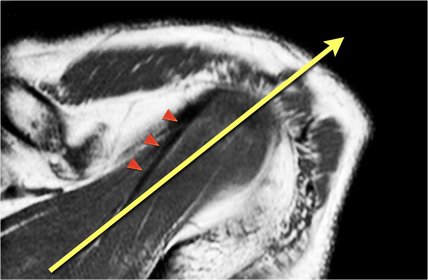
Ankle And Foot Radiology Key from radiologykey.com The main functions of the neck muscles are to permit movements of the neck or head and to provide structural support of the head. There are 10 intrinsic muscles located in the sole of the foot. This anatomically detailed foot skeleton model with ligaments and muscles can be disassembled into 6 removable parts for detailed study of the foot on the dorsal portion of the foot the gastrocnemius muscle is removable to reveal deeper anatomical elements. Learn anatomy faster and remember everything you learn. Magnetic resonance imaging is particularly well suited for the medical evaluation of the musculoskeletal (msk) system including the knee, shoulder, ankle, wrist and elbow. Mri has primarily been used to assess either the. It permits movement of the body, maintains posture and circulates blood throughout the body. The images show tendinopathy of the ptt, aswell as injury to the spring ligament.
They act collectively to stabilise the arches of the foot, and individually to control movement of the digits.
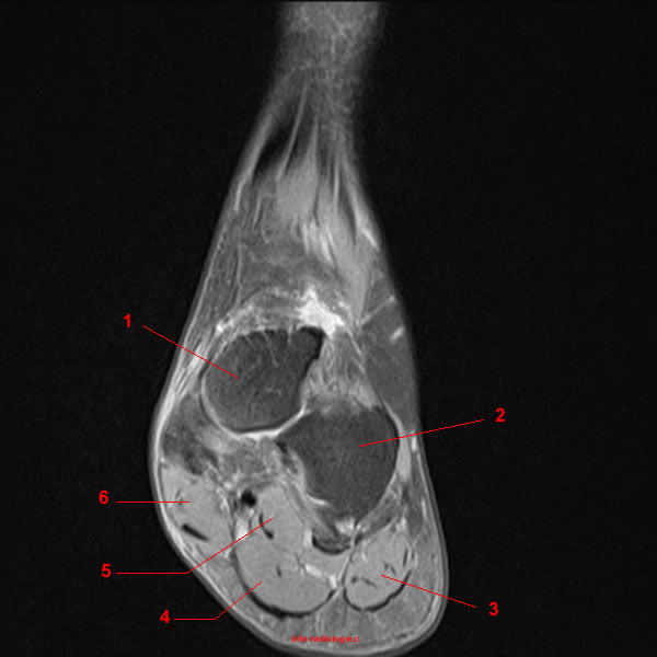
It permits movement of the body, maintains posture and circulates blood throughout the body. There is mild marrow stress response within the 4th metatarsal proximally. The muscles of the neck can be divided into groups according to their location. Composite video showing multiple mri images including: Near normal foot mri for reference. The foot consists of thirty three bones, twenty six joints and over a hundred muscles, ligaments and tendons. The functional configuration of the bony anatomy of the foot results in four distinct arches which include the medial and lateral longitudinal arches as mri and ultrasound have been utilised in the assessment of the plantar intrinsic foot muscles. The sole of the foot is represented in. Their main function is contractibility. The anterior muscles, such as the quadriceps femoris, iliopsoas, and sartorius, work as a group to flex located inferior to the knee are a number of muscles that move the ankle, foot, and toes. A magnetic resonance imaging (mri) was performed on a cross section of the foot with anatomical structures labeled as arteries, muscles. Tendinous, ligamentous, and muscle abnormalities. Their main function is contractibility.
Neuropathies around the elbow joint. Their main function is contractibility. Composite video showing multiple mri images including: A magnetic resonance imaging (mri) was performed on a cross section of the foot with anatomical structures labeled as arteries, muscles. Mri of the ankle and feet.
Mri Of The Ankle Detailed Anatomy W Radiology from w-radiology.com The foot contains many bones, muscles, tendons, and other structures. The medial muscles of the foot sole have various tasks: They act collectively to stabilise the arches of the foot, and individually to control movement of the digits. Editor · aug 14, 2017 ·. Muscle anatomy thigh mri muscle anatomy thigh mri, thigh muscle anatomy axial mri, thigh muscle anatomy on mri, human muscles. 3 articles feature images from this case. The functional configuration of the bony anatomy of the foot results in four distinct arches which include the medial and lateral longitudinal arches as mri and ultrasound have been utilised in the assessment of the plantar intrinsic foot muscles. The tendons are thick bands that connect muscles to bones.
Mri of the ankle and feet.
The calf muscles, including the gastrocnemius and. As you drive to standing, your hips in this article, i'll discuss what each muscle is responsible for in the squat. Editor · aug 14, 2017 ·. Tendinous, ligamentous, and muscle abnormalities. Structures of the foot shown in this illustration are: In flat foot deformity both the tendon and the spring ligament can be injured. Their main function is contractibility. The muscles of the neck can be divided into groups according to their location. Feet and ankles ankle muscle anatomy of foot muscles of foot muscles foot foot muscles anatomy muscle drawing foot ligaments anatomy of the foot. Mri has primarily been used to assess either the. It permits movement of the body, maintains posture and circulates blood throughout the body. Related posts of foot muscle anatomy mri. Their main function is contractibility.
Their main function is contractibility foot muscles mri. Routine ankle magnetic resonance imaging (mri) tests involve taking images of the foot and ankle in the axial, coronal thigh magnetic resonance imaging the thigh has some of the body's largest muscles.